No More “Take Two” in Lung Cancer Surgeries

Suffering from lung cancer and undergoing an operation is distressing enough. Yet some patients need to go through a second surgery when the tumor has not been completely removed. A HKUST research team has developed a cutting-edge microscope that can accurately detect any remaining cancer cells in just three minutes. Leveraging artificial intelligence, this innovation can help doctors make near real-time judgments during surgery while sparing patients from unnecessary ordeal.
Fighting a top killer
“Lung cancer is the leading cause of cancer mortality, with an estimated 2.2 million new cases and 1.8 million deaths globally in 2020. In general, in about 10% to 20% of surgery cases, patients need to return and undergo a secondary operation,” Prof. Terence WONG Tsz-Wai, the research project’s principal investigator and an assistant professor of the Department of Chemical and Biological Engineering, explains why the study is currently focusing on this disease.
“A major problem plaguing surgeons for many years is not knowing whether they have successfully excised all the cancer cells from the patients before leaving the operating theater.”
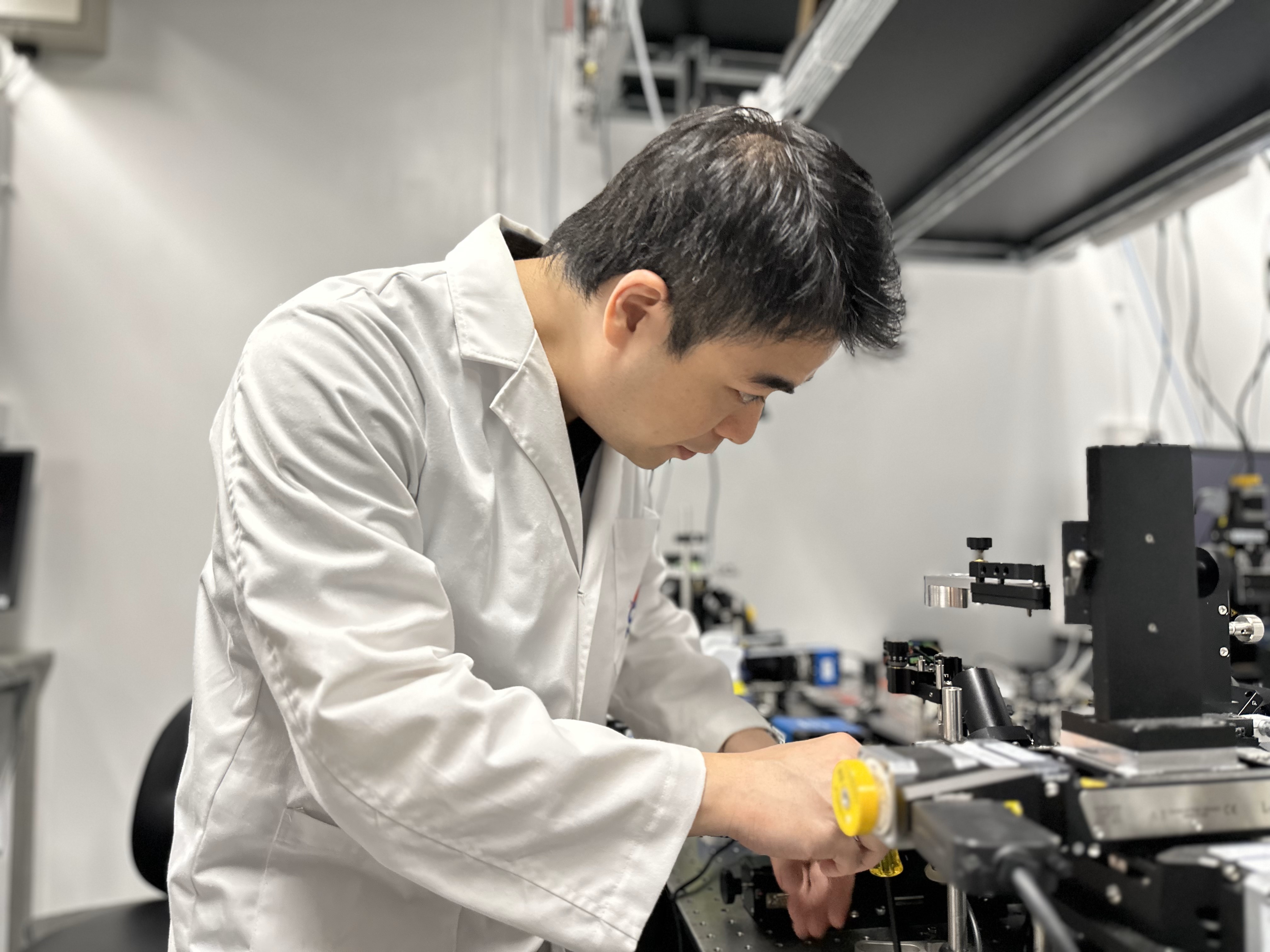
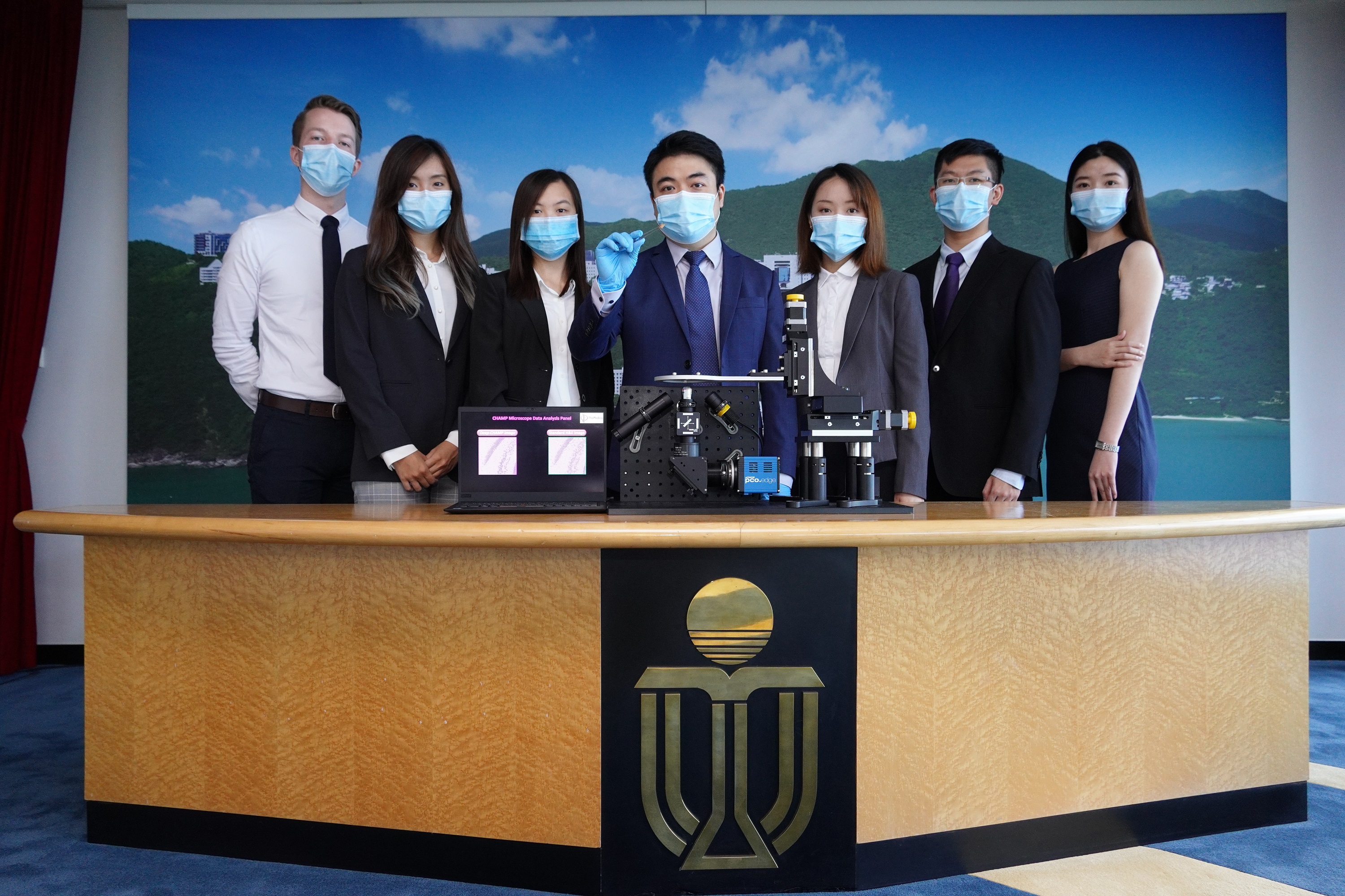
Prof. Terence Wong, pictured here working with the CHAMP microscope, aims to alleviate the suffering of cancer patients with his breakthrough in medical imaging technology.
Prof. Wong and his team are excitedly looking forward to seeing their innovation help patients very soon. Joining hands with five hospitals, they are conducting a large-scale clinical trial that covers about 1,000 patients’ tissue samples, with the microscope expected to be officially in service from 2024 onward locally and from 2025 onward on the Mainland.
Their collaborators are Queen Mary Hospital and Prince of Wales Hospital in Hong Kong, and three hospitals on the Mainland — Peking University Shenzhen Hospital, the People’s Hospital of Guangxi Zhuang Autonomous Region, and Anyang Tumor Hospital.
Solution for doctors, new hope for patients
Currently, there are two commonly used methods of cancer tissue imaging. Using a conventional microscope with a lengthy sample preparation procedure, widely regarded as the gold standard, has a near-perfect accuracy. However, it typically takes a week to generate results. This means patients will have to wait anxiously for a week to know whether the operation is successful. If not, they will have to suffer from a second surgery if they want to clear out the remaining cancer cells from their body.

Prof. Terence Wong (fourth from left) and his team pose for a photo at the Hong Kong Science Park, where their start-up’s office is situated.
Another option, known as the frozen section, can quickly generate results in 30 minutes. However, its accuracy is only about 70%.
The breakthrough in HKUST’s imaging technology enables the detection of cancer cells in just three minutes, with an accuracy of higher than 90% — comparable to the gold standard of the one-week conventional test.
“This is achieved by using ultraviolet (UV) light excitation to image the surface of the tissue at a specific wavelength,” Prof. Wong elaborates on the science behind his pioneering microscope.
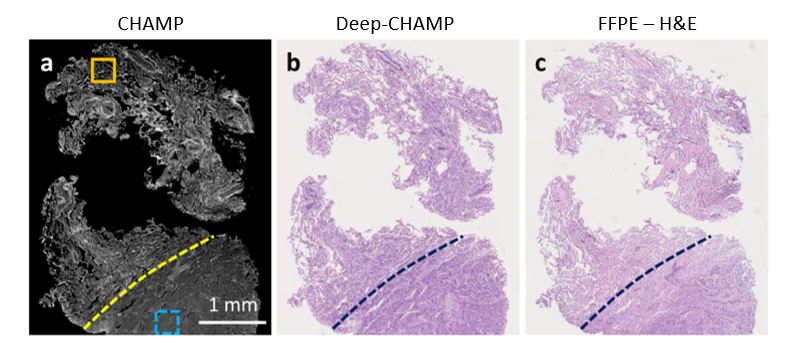
In just three minutes, the CHAMP microscope first generates a greyscale image (left, a) of the patient’s tissue sample and then virtually colors it (middle, b) to indicate the locations of cancer cells for doctors. The result is comparable to that of an image generated by a conventional one-week test (right, c), which is regarded as the gold standard in the medical profession.
“After a greyscale image is generated, a deep learning algorithm we developed will then transform it into a histological image convenient for doctors’ instant interpretation, enabling them to make sure all cancer cells have been cut in the operation.”
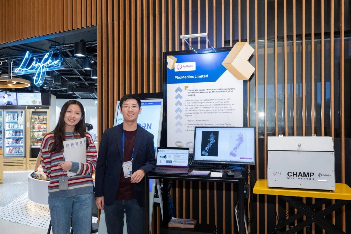
The technology, called Computational High-throughput Autofluorescence Microscopy by Pattern Illumination, or CHAMP in short, has been acclaimed locally, regionally, and internationally. With HKUST’s support, the team has launched a start-up called PhoMedics with funding from the Technology Start-up Support Scheme for Universities (TSSSU), aiming to translate research into applications in hospitals and clinics.
HKUST PhD student Victor Tsang, a co-founder of the start-up PhoMedics, presents the CHAMP technology at the 48th International Exhibition of Inventions Geneva. The team is honored with a gold medal at the event.
In 2020, the company was acclaimed with the Gold Award at the HKUST-Sino One Million Dollar Entrepreneurship Competition, and selected as the winner of the Innovation Nanshan Entrepreneurship Star Contest’s foreign channel, one of the largest start-up competitions in Shenzhen. This year, it was further applauded in the international science arena by winning a gold medal at the 48th International Exhibition of Inventions Geneva.
Apart from developing the technology, the company also plans to produce the CHAMP microscope and make it available to medical institutions in Hong Kong, the Mainland, as well as in overseas markets in the future.
The team led by Prof. Terence Wong (middle) is acclaimed with the Gold Award at the HKUST-Sino One Million Dollar Entrepreneurship Competition 2020.
Future of medical imaging
The invention is a culmination of years of meticulous research. During his PhD training at Washington University in St. Louis and the California Institute of Technology, Prof. Wong dedicated his efforts to the improvement of histological imaging. Under the tutelage of biomedical imaging expert Prof. WANG Lihong, he and his lab-mates successfully developed a microscope that could analyze breast cancer tumors in one to two hours with comparable accuracy as the gold standard of the one-week conventional test.
“The results were highly encouraging, but it was not fast enough. There should be room for further improvement. I was then determined to continue with my expedition in medical imaging research. After graduating with my PhD degree in the US, I returned to Hong Kong and joined HKUST as a full-time faculty member,” Prof. Wong says.
“Later, a pulmonologist approached me and asked if the technology could be applied to lung cancer. Since the lung is a vital organ, precise cutting during surgery would be critical to maximizing the preservation of patients’ respiratory function while minimizing the health risks posed by any remaining cancer cells. So, I took up the challenge and shifted the focus of my research to lung cancer. At the same time, as a principal investigator, I led my research team at HKUST to successfully develop CHAMP microscopy that is about 100 times faster than what I have developed during my PhD training, finally making this novel CHAMP microscope clinically useful and impactful.”
Besides lung and breast cancers, the potential applications of CHAMP are vast. The team is also conducting tests on smaller scales on liver, colorectal, kidney, and skin cancers, as well as prostate gland conditions.
“In the future, we are confident that the CHAMP technology will take medical imaging and diagnosis to a new level. Our microscope will serve as a useful device not only in Hong Kong hospitals but also nationwide and overseas,” Prof. Wong says.
(This news was originally published by the HKUST Global Engagement and Communications Office here.)